Recommendations for the use of structural magnetic resonance imaging in the care of patients with epilepsy: A consensus report from the International League Against Epilepsy Neuroimaging Task Force. Epilepsia. 2019
ILAEによるMRI精査コンセンサスレポート (Epilepsia 2019)
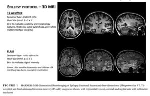
HARNESS-MRIプロトコールを推奨
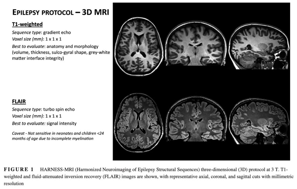
T1とFLAIRは3D MRIを推奨
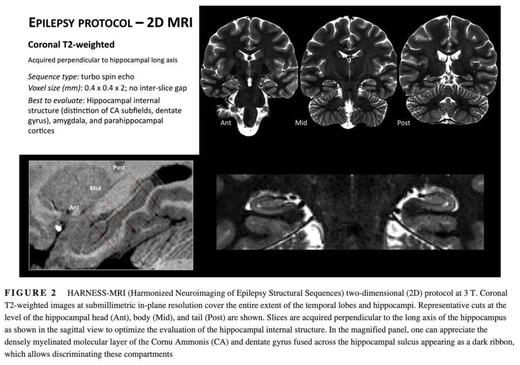
T2で海馬構造と周辺を確認

FLAIR画像. 3mmスライスだと見落とす
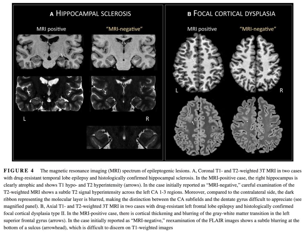
Aは2例の海馬硬化症を示している.
MRI negativeのケースも良く見ると, 左海馬構造に微小な高信号と海馬層構造不明瞭化を認める.
Bは両方ともFCD type Ⅱ.
MRI negativeとされたケースも微細なFCDがあることを示している.
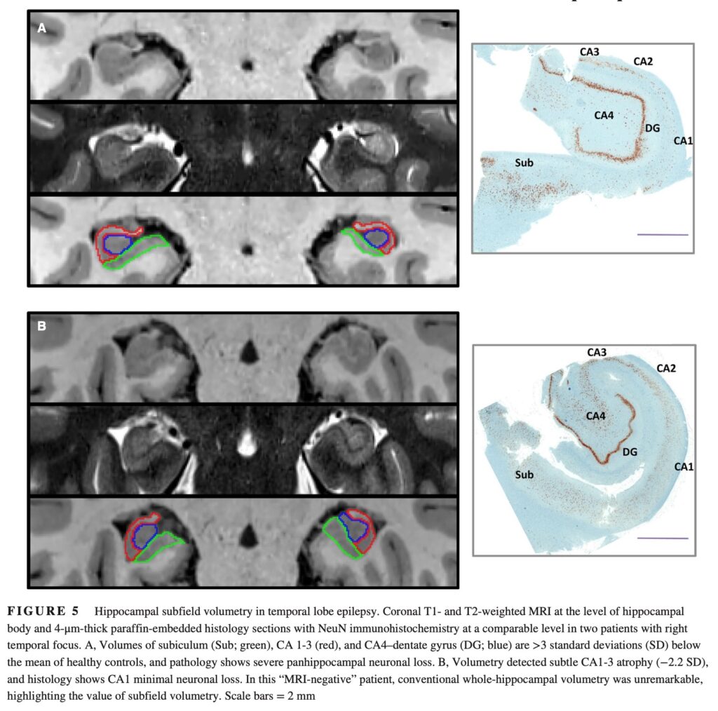
BはMRI negativeとされたが, Subfield volumetryによる評価で左CA1-3にわずかな萎縮を検出.
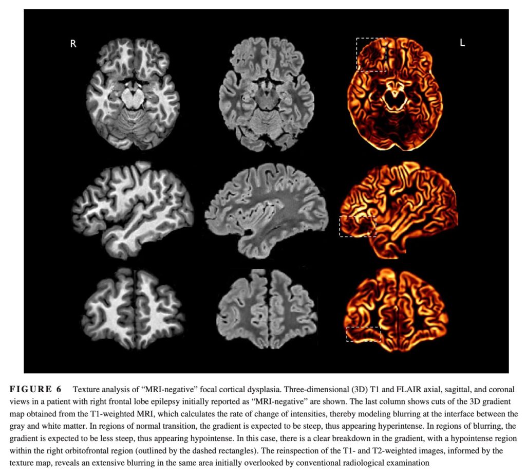
MRI negativeとされたケース.
T1, FLAIRから得られる灰白質のぼやけをtexture analysisで検出
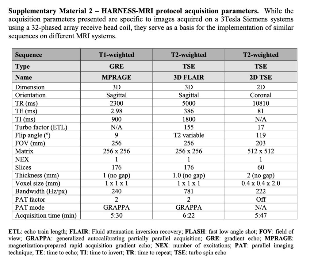
HARNESS-MRIプロトコールの詳細
[Bernasconi A, Cendes F, Theodore WH, et al. Recommendations for the use of structural magnetic resonance imaging in the care of patients with epilepsy: A consensus report from the International League Against Epilepsy Neuroimaging Task Force. Epilepsia. 2019;60(6):1054-1068. doi:10.1111/epi.15612]
【まとめ】
3D-T1, 3D-FLAIR, T2 (冠状断)は必須である.
FLAIRで3 mmスライスの場合は, FCDを見落とすことがある.
T2で海馬層構造の確認が必要である.
腫瘍, 血管奇形, 感染が疑われる場合は, SWI, T2*, 造影T1を追加する.
コメント